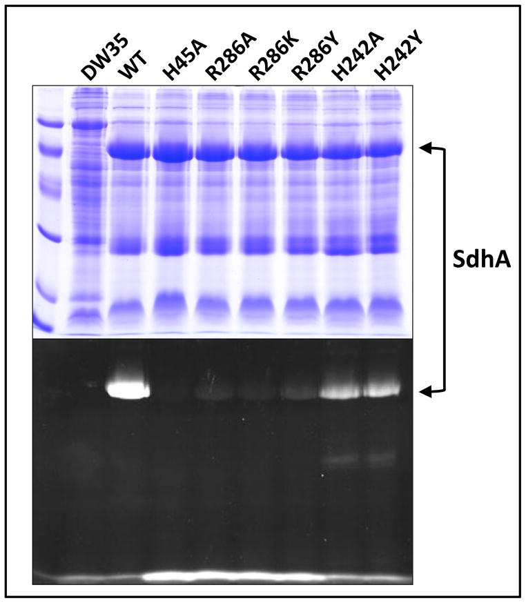
Figure 2. Enzyme assembly and covalent FAD incorporation.
Proteins were separated on a 12 % SDS-PAGE gel and visualized by either Coomassie Blue staining (top) or UV excitation (bottom). Samples from left to right: low molecular weight standards, followed by enriched membrane preparations containing no Sdh (E. coli DW35), wild-type Sdh (WT), SdhA-H45A, -R286A, -R286K, -R286Y, -H242A and -H242Y enzymes.

