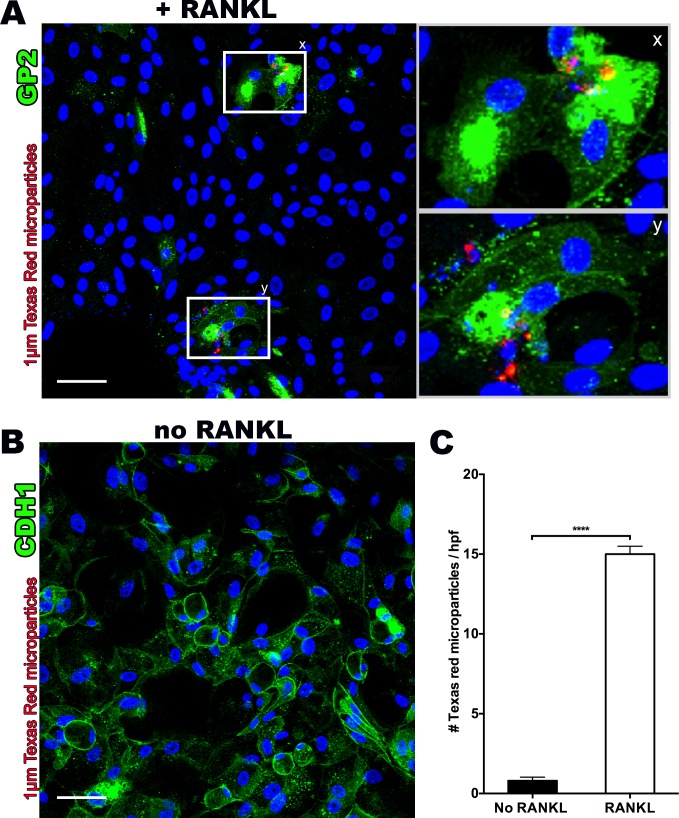
Fig 4. RANKL treated monolayers demonstrate functional M cells through uptake of fluorescent 1μm Texas Red latex microparticles.
(A) Confocal microscopy z-stack assessment (400X magnification) showing colocalization of fluorescent 1μm Texas Red latex microparticles and GP2 positive stained M cells with enlarged sections [x,y] showing clumping of microparticles. (B) Confocal microscopy section (400X magnification) of non-RANKL monolayer stained for E-cadherin (CDH1), a cell surface marker that M cells lack, demonstrating uniform and ubiquitous staining, as well as lack of any Texas Red microparticle internalization. (C) RANKL treated monolayers show significantly increased numbers of retained fluorescent microparticles after washing, as assessed by three random high power field areas under confocal microscopy z-stack projections (400x). Scale bar 50 μm. ****p<0.0001.

