
Fig 1. Intracellular expression and extracellular secretion of MANF and CDNF in HEK293 cells.
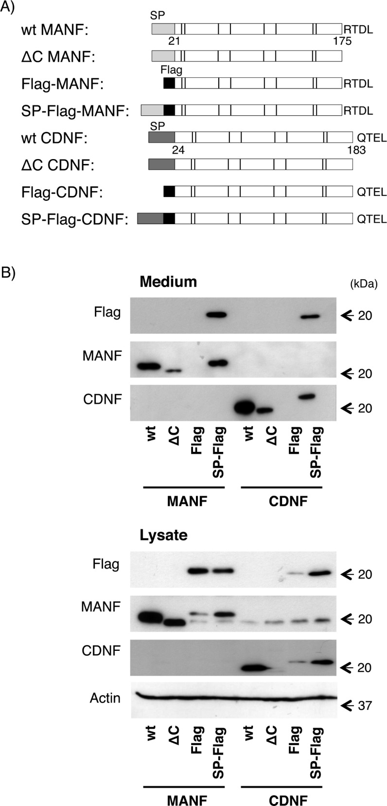
(A) Schematic representation of the mouse MANF and CDNF expression constructs used in this study. SP indicates a signal peptide at the N-terminus of each protein. The cysteines are indicated by bars. The four C-terminal amino acids, RTDL and QTEL, putative ER localization signals at their C-termini are shown in capital letters. (B) Western blot analysis of wild-type and modified MANF and CDNF overexpressed in HEK293 cells. Twenty-four hours after transfection of each indicated construct into the cells, the culture medium was replaced with fresh serum-free DMEM, and the cells were incubated for an additional 12 h. The amounts of the indicated proteins in the cell lysate and culture medium were detected by western blot analysis using antibodies against Flag-epitope, MANF, CDNF and actin as described in the Materials and Methods. Representative data of three independent experiments were shown.