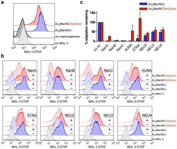
Figure 3. Activity of sialidases toward α2-3-linked sialic acids on BJAB K20 cell surfaces.
(a) α2-3-linked sialic acids on the surface of BJAB K20 cells were detected by flow cytometry using MAL II lectin. Cells cultured with no mannosamine precursor do not produce α2-3-linked sialic acids. (b) BJAB K20 cells were cultured with Ac4ManNAc, Ac4ManNDAz(2me), or no mannosamine, then left untreated or treated with the indicated sialidase. Remaining α2-3-linked sialic acids on the cell surface were detected by flow cytometry using MAL II lectin. (c) Quantification of analyses shown in panel (b), representing the amount of α2-3-linked Neu5Ac or SiaDAz(2me) remaining on BJAB K20 cell surfaces after treatment with the indicated sialidase. Error bars represent the standard deviation of three trials.

