Figure 2.
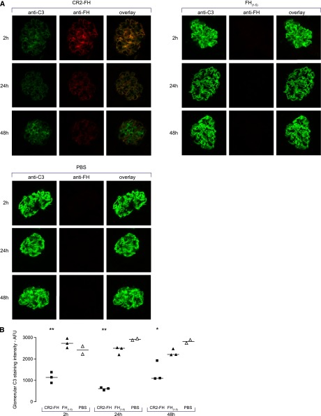
CR2-FH colocalized with and reduced glomerular C3 in Cfh−/− mice. (A) Representative images of glomerular C3, CR2-FH, and FH(1–5) in Cfh−/− mice injected with a single dose of CR2-FH, FH(1–5), or PBS. C3 was visualized using FITC-conjugated goat anti-mouse C3 polyclonal antibody (green). Alexa 594-conjugated rabbit anti-mouse FH polyclonal antibody was used to detect either CR2-FH or FH(1–5) (red). CR2-FH (red) colocalized with C3 (green) along the GBM. No staining was observed with the Alexa 594-conjugated rabbit anti-mouse FH polyclonal antibody in mice injected with either FH(1–5) or PBS. Granular C3 was detected 48 hours after CR2-FH injection. Original magnification, ×40. (B) Quantitative immunofluorescence demonstrated a reduction in glomerular C3 in the CR2-FH group compared with either the FH(1–5) or PBS-injected mice at 2, 24, and 48 hours. Horizontal bars denote median values. **P<0.001 versus FH(1–5) and P<0.01 versus PBS. *P<0.01 versus FH(1–5) and P<0.001 versus PBS, Bonferroni’s Multiple Comparison Test. AFU, arbitrary fluorescent units.

