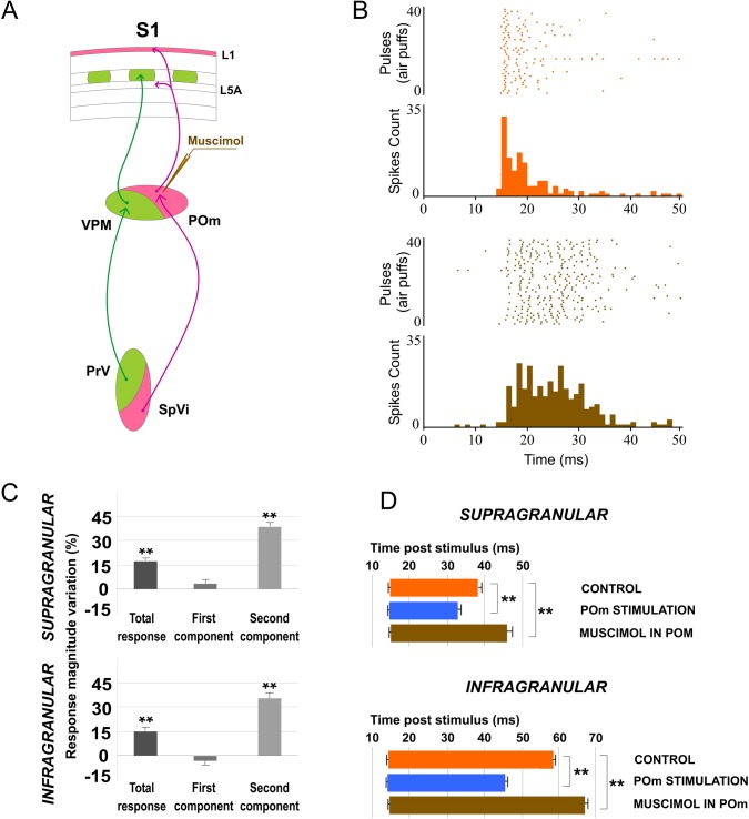
Fig 4. Muscimol-induced inactivation of the POm.
(A) Schematic diagram indicating the experimental manipulation of the paralemniscal (pink) thalamocortical circuitry to barrel cortex. (B) POm inactivation enhanced responses in S1 mainly in the second component. Raster plots and PSTHs are shown for a sample supragranular neuron before (top) and after (bottom) POm inactivation. Also the pattern of spikes in the response was changed after POm inactivation suggesting that POm imposes a precise control of cortical responses. (C) Percentage change in mean response magnitude when POm was inactivated with muscimol. Spikes were strongly enhanced in the second component of the response. (D) Mean onset and offset latencies and response duration in Control (orange), in POm E-stimulation (blue) and in POm inactivation condition (brown). Response duration decreased with POm E-stimulation and increased in POm inactivation condition. We did not find differences in onset latencies but offset latencies changed significantly. Horizontal bars represent response duration.

