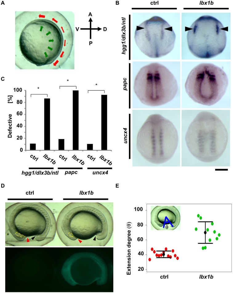
Fig 5. Defective convergent extension in lbx1b overexpressing zebrafish embryos.
(A) A diagram of convergent extension during zebrafish gastrulation. A, P, D, and V indicate anterior, posterior, dorsal, and ventral sides, respectively. The green and red arrows indicate the directions of convergence and extension movements, respectively. (B) In situ hybridization for germ layer markers at the early stages of development. Dorsal views of embryos injected with buffer as control (ctrl) or lbx1b mRNA (lbx1b). Upper panels show marker expression of hgg1 (rostral mesoderm), ntl (notochord), and dlx3b (border between neural and non-neural ectoderm, black arrowheads) at the tail bud stage. Middle panels show the expression of papc (paraxial mesoderm) at 11 hpf. Lower panels show the expression of uncx4 (somite) at 13 hpf. The scale bar represents 200 μm. (C) Incidence of defective expression patterns. Significant differences (*p < 0.01) were observed for the germ layer markers. The numbers of control and lbx1b zebrafish embryos were 36 and 30 for hgg1/ntl/dlx3b, 32 and 28 for papc, and 28 and 27 for uncx4, respectively. (D) Lateral views of 16 hpf normal sibling (ctrl) and Tg(hsp:Gal4-VP16:EGFP:UAS:lbx1b) (lbx1b) embryos upon heat shock at 4 hpf. The red and black arrowheads indicate the border between the yolk and the rostral part and the yolk and the caudal part, respectively. The yellow lines indicate the developing eyes. (E) Quantitative analysis of extension movement. Progression of the movement was quantified by the angle θ at 16 hpf for sibling control (ctrl; n = 13) and transgenic (lbx1b; n = 11) embryos. Convergent extension movement was significantly delayed in lbx1b embryos (p < 0.01).

