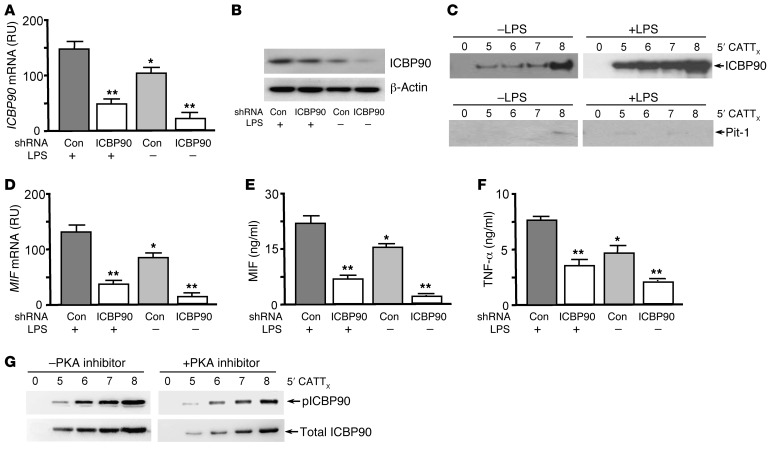
Figure 3. ICBP90 binds to the MIF –794 CATT5–8 promoter sequence in an activation- and CATT length–dependent manner.
(A) ICBP90 mRNA expression analyzed by qPCR in human THP-1 monocytes (1 × 10⁶) treated with 0.5 μg of a control or ICBP90 shRNA prior to stimulation with LPS (100 ng/ml, 16 hours). (B) Corresponding cellular content of ICBP90, as measured by Western blotting, with anti-ICBP90 or anti–β-actin as a protein control. (C) Detection of DNA-bound ICBP90 after incubation of the nuclear lysates that were collected from the cells used to determine cellular ICBP90 content with 100 nM each of the biotin-labeled 5′CATT0–8 oligonucleotides spanning the MIF promoter (–865/–833 to –752). The 5′CATT0–8 oligonucleotide–bound proteins were removed by streptavidin bead absorption after 3 hours at 4°C, and 1 μg of each sample was electrophoresed and immunoblotted with an anti-ICBP90 or an anti–Pit-1 antibody. (D) Quantification by qPCR of MIF mRNA in human THP-1 monocytes treated with ICBP90 or control shRNA followed by stimulation with LPS (100 ng/ml, 16 hours), together with corresponding effects on (E) MIF and (F) TNF-α protein secretion into 24-hour-conditioned medium. (G) Western blot detection of 5′CATT0–8-bound ICBP90 after incubation of nuclear lysates from LPS-stimulated human THP-1 monocytes cotreated with the PKA inhibitor H-89 (20 μM) or vehicle control. The 5′CATT0–8 oligonucleotide–bound proteins were removed by streptavidin bead absorption after 3 hours at 4°C and 1 μg of each sample was electrophoresed and immunoblotted with an anti-ICBP90 (Total) or anti-pICBP90 antibody. Data are mean+SD of 3 measurements, with all experiments replicated twice (n = 3 measurements per experiment). *P < 0.05 for LPS– vs. LPS+ controls, **P < 0.01 for ICBP90 shRNA vs. corresponding control shRNA (Con) (1-way ANOVA with Tukey’s post-hoc test).

