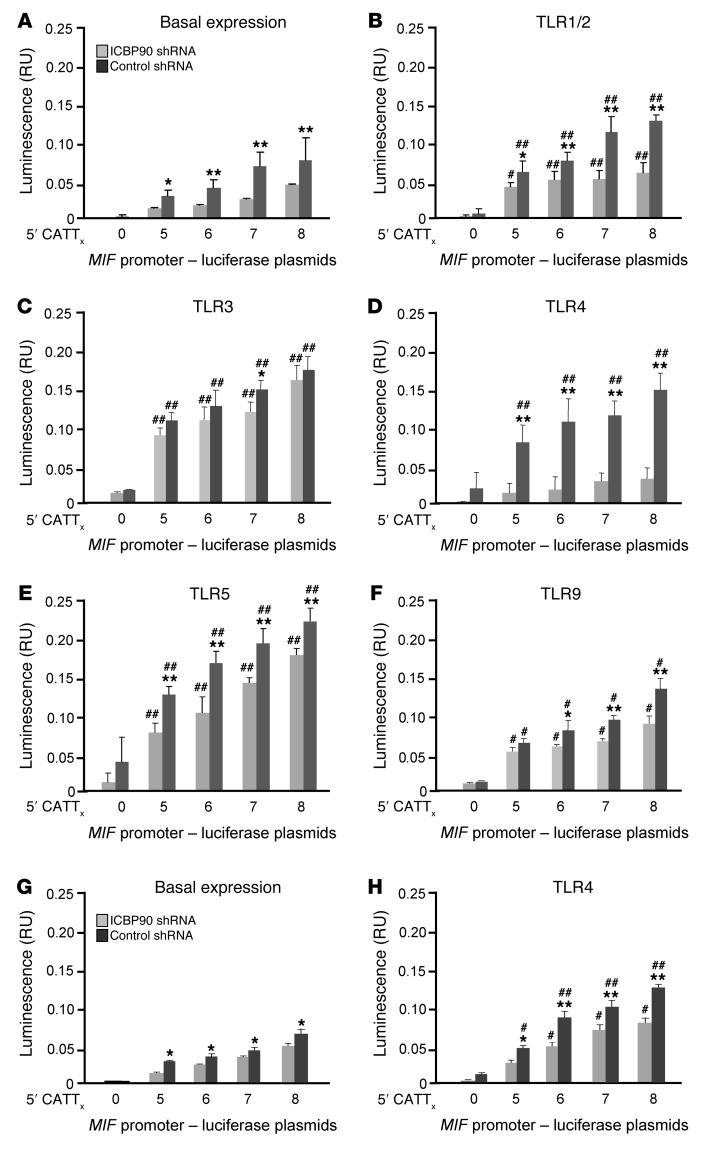
Figure 4. Regulation of TLR-activated MIF expression in human monocytes by ICBP90.
THP-1 monocytes were transfected with MIF promoter-luciferase reporter plasmids bearing 0, 5, 6, 7, and 8 CATT repeats and treated with an ICBP90 or control shRNA, cultured for 6 hours, and (A) were left unstimulated or were stimulated with (B) Pam3CysK (100 ng/ml) for TLR1/2 agonism, (C) polyI:C (1 μg/ml) for TLR3 agonism, (D) LPS (100 ng/ml) for TLR4 agonism, (E) flagellin (100 ng/ml) for TLR5 agonism, or (F) CpG DNA (5 μM) for TLR9 agonism prior to measurement of luciferase activity. Primary human peripheral blood monocytes were similarly analyzed under (G) basal and (H) LPS-stimulated conditions (100 ng/ml). Data are presented as the mean+SD of 3 measurements, with all experiments replicated twice (n = 3 measurements per experiment). *P < 0.05, **P < 0.01 for control shRNA vs. ICBP90 shRNA within each panel; #P < 0.05, ##P < 0.01 for stimulated vs. basal expression for B–F vs. A and for H vs. G (2-tailed Student’s t test).

