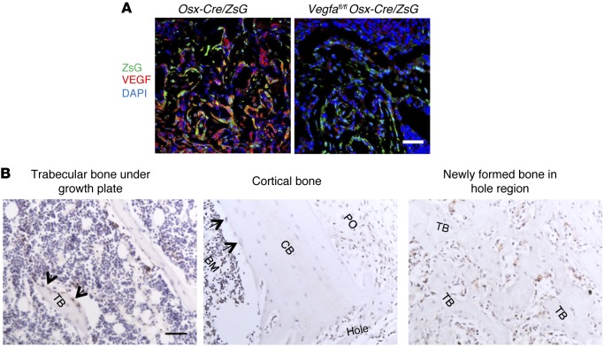
Figure 1. Osteoblastic lineage cells are an important source of VEGF at the bone-repair site.
(A) At PSD7, low density (6.1% ± 1.8%) of anti-VEGF staining (red) in defect hole region of Vegfafl/fl Osx-Cre/ZsG mice compared with Osx-Cre/ZsG mice (15.5% ± 2.1%); P < 0.05. The percentage of VEGF-expressing ZsG+ osteolineage cells (yellow) of total ZsG+ cells (yellow cells plus green cells) was 17.0% ± 5.2% in the hole region of Vegfafl/fl Osx-Cre/ZsG mice compared with Osx-Cre/ZsG mice (51.5% ± 8.3%); P < 0.05. Finally, the percentage of VEGF-expressing ZsG+ osteolineage cells (yellow) of total VEGF-expressing cells (yellow cells plus red cells) was also reduced in hole region of Vegfafl/fl Osx-Cre/ZsG mice (23.0% ± 1.8%) compared with Osx-Cre/ZsG mice (72.6% ± 6.2%); P < 0.05. (B) VEGF staining of trabecular bone, cortical bone, and the newly formed bone within cortical defects in WT mice at PSD7. Black arrows indicate VEGF-expressing osteoblasts lining the surface of trabecular bone (TB) or endosteum of cortical bone (CB). Representative images from 3 mice. Scale bar: 50 μm (A and B). Unpaired 2-tailed Student’s t test was used for comparisons of percentages between groups; n= 4–6 for each genotype.

