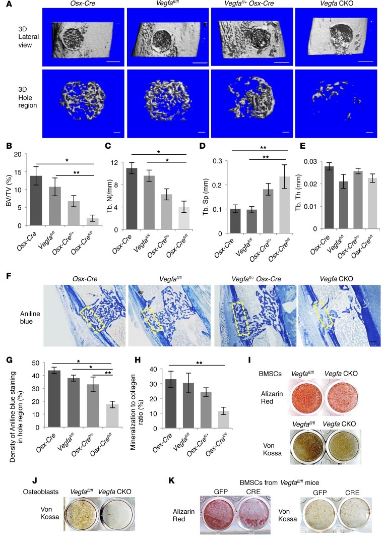
Figure 2. Impaired intramembranous bone formation in defects of Vegfa CKO mice at PSD7.
(A) Lateral views of 3D reconstruction of injured tibiae (top panel, scale bar: 500 μm) and mineralized bone formed in hole region (lower panel, scale bars: 100 μm) by μCT. Representative images from 6 mice of each genotype. (B–E) 3D structural parameters — trabecular BV/TV%, Tb.N, Tb.Sp, and Tb.Th — of mineralized bone formed in hole region by μCT; n = 6. (F) Aniline blue staining shows reduced collagen accumulation in hole region of Vegfa CKO mice. Density of aniline blue staining calculated for hole region (stippled yellow rectangles). Scale bar: 200 μm. (G) Low density of aniline blue–stained area in hole region of Vegfa CKO mice; n = 8–11. (H) Decreased mineralization/collagen ratio in Vegfa CKO mice; n = 5–6. (I and J) Alizarin red and Von Kossa staining of BMSCs (I) and osteoblasts (J) after culture in mineralization medium containing 100 nM dexamethasone for 21 days. (K) Alizarin red and Von Kossa staining of BMSCs (from Vegfafl/fl mice) treated with adenoviral GFP or Cre and cultured in mineralization medium with 100 nM dexamethasone for 21 days. The data are representative of 3 independent experiments (I–K). ANOVA with Tukey’s post-hoc test was used. *P < 0.01; **P < 0.05.

