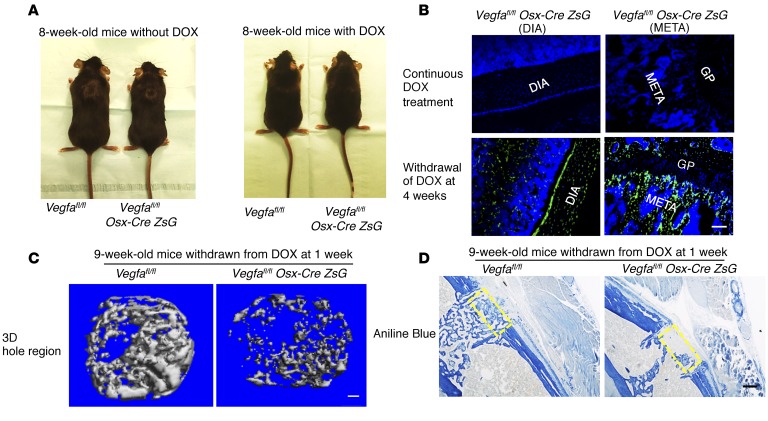
Figure 4. Postnatal deletion of Vegfa in osteoblast lineage cells impairs intramembranous bone formation in cortical defects.
(A) Reduced body size and weight in 8-week-old Vegfa CKO mice (18.4 ± 0.7 g) compared with Vegfafl/fl mice without doxycycline (DOX) treatment (23.4 ± 0.7 g); n = 8–10, P < 0.01. This reduction was eliminated in DOX-treated mice (20.8 ± 0.3 g and 21.7 ± 1.1 g in the 2 groups); n = 4–6. (B) Absence of Cre-activated ZsG in diaphysis (DIA) and metaphysis (META) of 8-week-old Vegfafl/fl Osx-Cre/ZsG mice continuously fed with DOX; ZsG induced in mice by DOX withdrawal at 4 weeks. GP, growth plate. Representative images from 3 mice for each genotype. (C) μCT analysis of mineralized bone formed in hole region of 9-week-old Vegfa CKO mice with DOX withdrawn at 1 week shows reduced BV as percentage of total volume (BV/TV, 5.2% ± 1.9%), reduced Tb.N (5.4/mm ± 0.7/mm), but increased Tb.Sp (0.21 ± 0.03 mm) compared with that of Vegfafl/fl mice (BV/TV, 19.7% ± 2.4%; Tb. N, 11.9/mm ± 1.2/mm; Tb. Sp, 0.09 ± 0.01 mm). No significant differences were seen in Tb.Th; n= 6–10 for each genotype. BV/TV and Tb.N, P < 0.01; Tb.Sp, P < 0.05. (D) Reduced density of aniline blue staining (19.5% ± 4.2%) and decreased mineralization/collagen ratio (15.6% ± 5.1%) in hole region of 9-week-old Vegfa CKO mice with DOX withdrawn at 1 week, compared with Vegfafl/fl mice (40.8% ± 1.9% and 43.6% ± 7.2%); n= 6–10 for each genotype. Aniline blue staining, P < 0.01; mineralization/collagen ratio, P < 0.05. Yellow stippled rectangle: hole region. Scale bars: 100 μm (B and C), 200 μm (D). Unpaired 2-tailed Student’s t test was used for comparisons in C and D.

