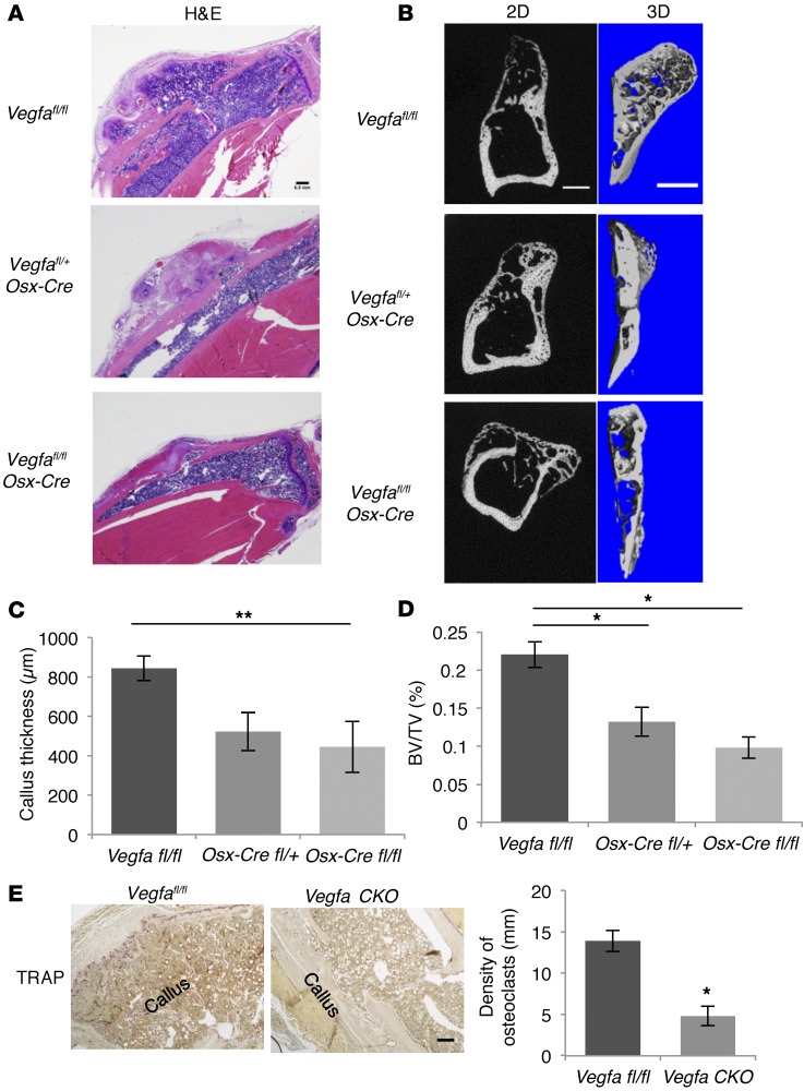
Figure 8. Deletion of Vegfa in osteoblastic cells reduces periosteal callus remodeling at PSD28.
(A) H&E-stained sections from Vegfafl/fl, Vegfafl/+ Osx-Cre, and Vegfa CKO mice showing periosteal callus remodeling at PSD28. Representative images from 5–6 mice for each genotype. (B) Left panels: 2D images of sagittal sections of injured tibiae. Right panels: 3D reconstruction of periosteal callus. Representative images from 5–6 mice. (C) Decreased callus thickness, calculated as distance from edge of periosteal callus to injured cortical bone normalized to total volume of corresponding tibial segment, in Vegfa CKO compared with Vegfafl/fl mice; n = 5–6. (D) Decreased BV/TV, total callus BV normalized to total volume of the corresponding tibial segment, in Vegfa CKO compared with Vegfafl/fl mice; n= 5–6. (E) Low density of TRAP+ osteoclasts, normalized to total length of callus bone, in Vegfa CKO compared with Vegfafl/fl mice; n = 4. Scale bars: 500 μm (A and B), 200 μm (E). ANOVA with Tukey’s post-hoc test (C and D) and unpaired 2-tailed Student’s t test (E) were used. *P < 0.01; **P < 0.05.

