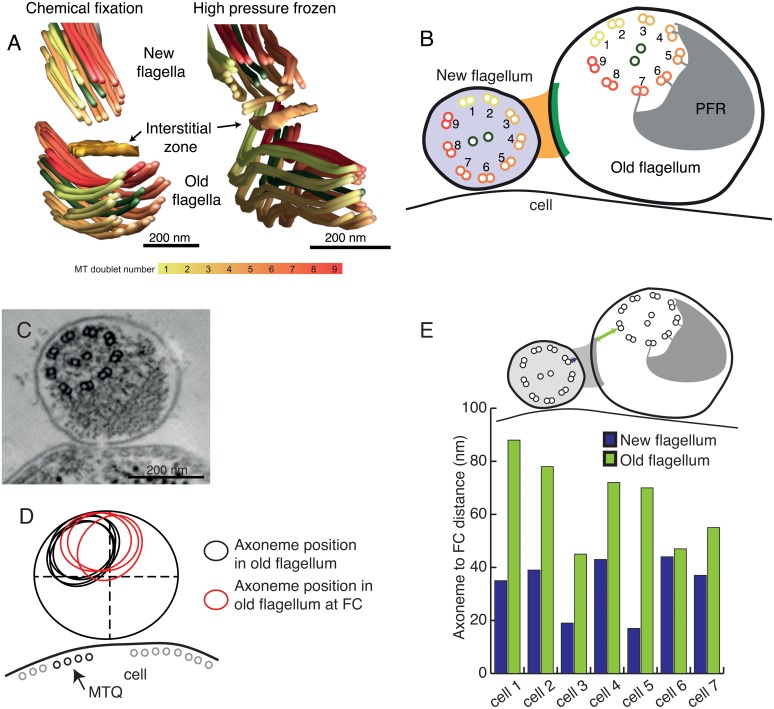
Fig 5. The FC displaces the old flagellum axoneme and its position is fixed in relation to both axonemes.
A) 3D reconstructions of the FC region show it in close proximity to microtubule doublets 3–5 in the new flagellum and microtubule doublets 1, 7–9 in the old flagellum both in high pressure frozen and chemically fixed samples. B) A cartoon illustrating the FC and the axoneme orientations in cross-section. C) An example tomographic slice of a 30 nm thick flagellum cross-section, oriented with the microtubule quartet to the left, shows the axoneme located in the top-left corner of the flagellar space. D) The line drawing shows a few examples of axoneme positioning within the old flagellum (black ellipsoids), and at the flagella connector (red ellipsoids). E) The distances between the FC and the nearest doublet microtubule are longer in the old flagellum.

