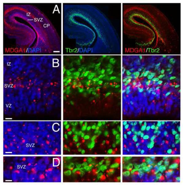
Figure 1. MDGA1 is expressed by basal progenitors of the subventricular zone.
Shown is immunolocalization of MDGA1 protein (red) and Tbr2 protein (green), a marker selective for basal progenitors of the subventricular zone (SVZ), on the same coronal cortical sections at E16.5. DAPI (blue) is shown as counterstaining. The C-D sets of photos are higher power views of SVZ cells from the A-B sets. Merged images of MDGA1 and Tbr2 are also shown. The majority of MDGA1+ cells in the SVZ (DM: 0.845±0.08; L: 0.32±0.01) co-localize with Tbr2 (tM: 0.54±0.06) and only a small fraction is negative for Tbr2. MDGA1 expression in the VZ describes a low-DM (0.154±0.07) to high-L (0.68±0.01) gradient. Abbreviations: CP: cortical plate; DM: dorsomedial; IZ: intermediate zone; L: lateral; VZ: ventricular zone. Scale bars: A (0.2 mm), B (100 μm) and C-D (50 μm).

