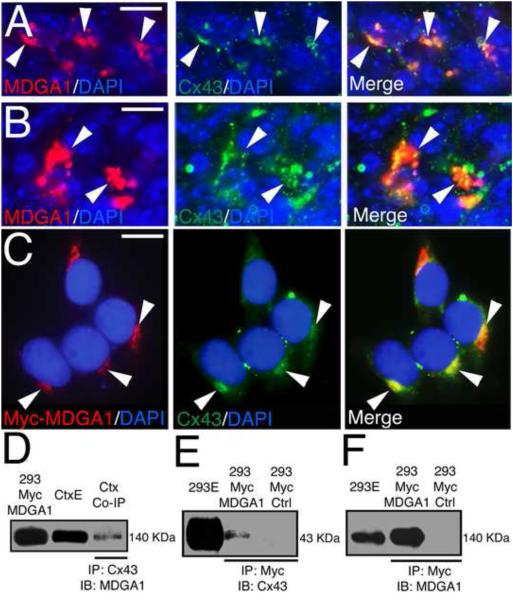
Figure 3. MDGA1 co-localizes and associates with the gap junction protein Connexin43.
Shown is co-localization of MDGA1 with Connexin43 (Cx43) in vivo in BPs of the SVZ (A-B) and in vitro in 293 cells transfected with Myc-MDGA1 (C), and the association of MDGA1 with Cx43 shown using co-immunoprecipitation (Co-IP) and immunoblotting (D-F). A-B: Immunofluorescence of MDGA1 (red) and Cx43 (green) on cortical sections at E16.5. Both MDGA1 and Cx43 show robust protein expression in the SVZ that is frequently co-localized in the plasma membrane of BPs in the SVZ. Arrowheads point to same cells in the respective series. C: 293 cells transfected with Myc-MDGA1 and immunostained for MDGA1 using a Myc antibody and for endogenous Cx43. Myc-MDGA1 (red) and the endogenous Cx43 (green) co-localize in discrete membrane domains. Arrowheads mark protein co-localization in the same domains in the series. Merged images are also shown. DAPI (blue) is used as counterstaining. D-F: Co-IP assays using protein extracts from WT cortex (E18, P7, D) or protein extracts of 293 cells transfected with Myc-MDGA1 (293 Myc MDGA1, E-F). D: Co-IP using WT cortical extracts (Ctx) and a Cx43 antibody, immunoblotted using an antibody specific for MDGA1 reveals a 140 KDa band as expected for MDGA1. E: Co-IP done with Myc antibody and immunoblot done with Cx43 antibody reveals a 43 KDa band as expected for Cx43. F: In a similar Co-IP, an immunoblot using an antibody specific for MDGA1 recognizes a 140 KDa band as expected for MDGA1. Abbreviations: 293E: whole extracts from 293 cells transfected with Myc-MDGA1; 293 Myc Ctrl: Myc negative control in 293 cells, no bands are labeled in the immunoblots; CtxE: whole cortex extract; IB: immunoblot; IP: immunoprecipitation. Scale bars: A-B (20 μm) and C (8 μm).

