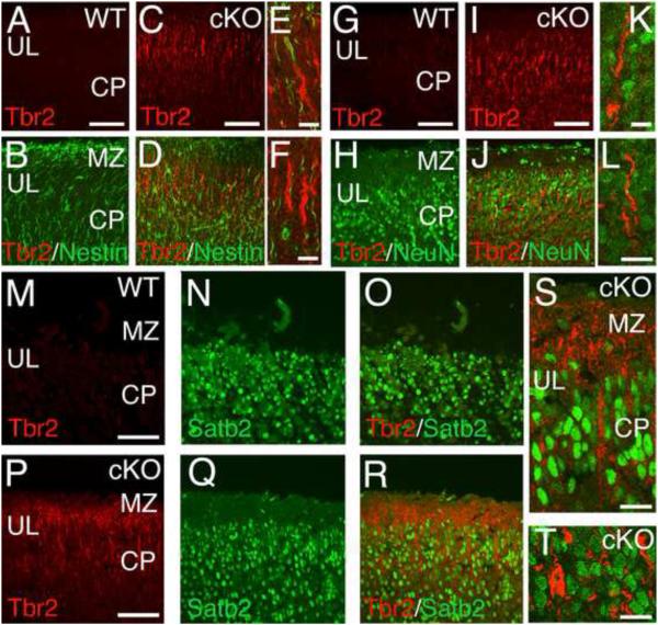
Figure 6. Ectopic Tbr2-positive cells in MDGA1 cKO cortex do not express neuronal or glial markers.
Shown are confocal images of sections from P0 WT and MDGA1 cKO cortex double immunostained with antibodies for Tbr2 (red), which in WT cortex is a marker specific for basal progenitors of the subventricular zone, and markers of radial glia (Nestin, green) and differentiated neurons (NeuN and Satb2, green), to determine possible cellular differentiation of the ectopic Tbr2+ cells found in the cortical plate (CP) of the cKO. The genotype and immunostaining for each panel is as indicated. Each panel shows only the upper layers (UL) of the CP and marginal zone (MZ). Ectopic Tbr2+ cells are found in the cKO cortex, but not in WT cortex; at this age (P0), they have a radial morphology and immunolocalization of Tbr2 protein (red) is confined to the apical process and cytoplasm of the cell body. The other markers (Nestin, NeuN and Satb2; green) are found in both WT and cKO cortex: in both, Nestin is localized to the radial processes of radial glia, which extend through the CP, whereas the transcription factors NeuN and Satb2 are localized to neuronal nuclei. Tbr2 immunostaining does not co-localize with immunostaining for any of the other three markers, as evident in the merged images from cKO cortex at low power (D) and high power (E,F) for Tbr2 and Nestin, at low power (J) and high power (K,L) for Tbr2 and NeuN, or at low power (R) and high power (S,T) for Tbr2 and Satb2. Scale bars: A-D (50 μm), E-F (20 μm), G-J (50 μm), K-L (20 μm), M-R (50 μm) and S-T (20 μm).

