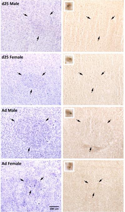
Figure 3.
Photographs of RA in males and females at 25 days of age (d25) and in adulthood (Ad). Pairs of nissl stained sections (left) and those exposed to immunohistochemistry for BDNF and BrdU (right) are adjacent. Arrows indicate the borders of the brain region. Inserts (right column) indicate cells labeled for both BrdU (blue/gray, nuclear) and BDNF (brown, cytoplasmic).

