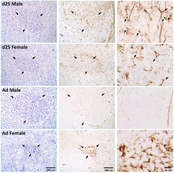
Figure 6.
Photographs of RA in males and females at 25 days of age (d25) and in adulthood (Ad). The nissl stained sections (left column) and those exposed to immunohistochemistry for vimentin and BrdU (middle column) are adjacent. Arrows in these sections indicate the borders of the brain region. The column on the right contains photos of higher magnification from near the center of RA in the immunohistochemical sections immediately to their left. Arrows indicate BrdU+ nuclei and vimentin+ fibers that appear to be in contact. Birds in the top two images (25-day-old) received scores of “3” in the qualitative assessment of contacts between BrdU− and vimentin-positive cells. The bottom two (adults) both received scores of “0”.

