
Fig. 11.
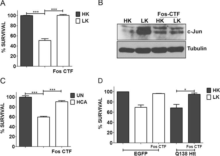
Fos-CTF is neuroprotective. a CGN cultures were treated with HK, LK, or LK+10 μM Fos-CTF for 24 h. c Cortical cultures were either left untreated (UN) or treated with 1 mM HCA with or without 10 μM Fos-CTF. Cell viability was assessed by performing Live-Dead assay 24 h and 18 h after treatment, respectively. b CGN cultures were treated with medium containing HK, LK, HK+10 μM Fos-CTF, or LK+10 μM Fos-CTF. Six hours after treatment, lysates were prepared and samples were subjected to Western blot analysis for c-Jun expression. Tubulin serves as a loading control. d CGNs were transfected with EGFP or Q138-Htt-GFP, 10 μM of Fos-CTF was added to the culture medium after transfection. Eight hours after transfection, cells were treated with HK, LK, HK+Fos-CTF, or LK+Fos-CTF medium. Immunocytochemistry was performed 24 h after the treatment and cell viability of transfected cells was assessed and normalized to EGFP transfected control. Results were obtained from at least three separate experiments done in duplicates. Statistical analysis using Student’s t test was performed where *** represents p<0.0001 and * represents p<0.05