
Fig. 2.
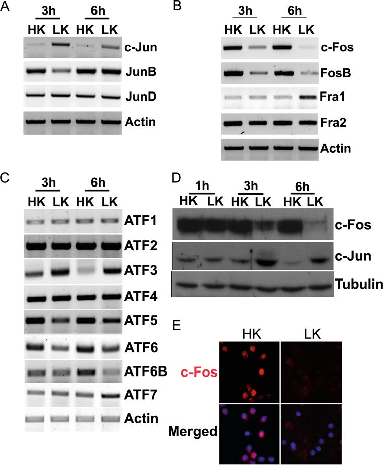
Expression profile of AP-1 members in postmitotic neurons. RNA isolated from CGNs treated with HK or LK for 3 and 6 h was subjected to RT-PCR analysis for Jun family (a), Fos family (b), and ATF family (c) expression. Actin serves as a loading control. d Whole-cell lysates prepared from CGNs treated with HK or LK for 1, 3, and 6 h were subjected to Western blot analysis using c-Fos and c-Jun antibody. Tubulin serves as a loading control RT-PCR, and Western blot were done at least three times from different samples. e CGNs were treated with HK or LK for 6 h. Immunocytochemistry with a c-Fos antibody was performed to visualize c-Fos levels. DAPI was used to visualize nuclei