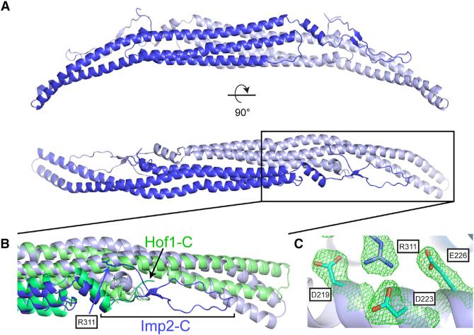
Figure 2. Structure of the Imp2 F-BAR Domain.
(A) Crystal structure of the Imp2 F-BAR domain dimer with subunits colored in dark and light blue.
(B) Close-up view of the wing-tip orientation and C-terminal residues of Imp2 (blue), which diverge significantly from Hof1 (green).
(C) Close-up view of the intermolecular salt-bridge network formed by the F-BAR C terminus (2Fo – Fc contoured at 1.1σ).

