Figure 3.

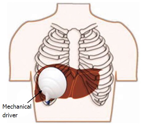
Typical transcostal position of the passive driver for magnetic resonance elastography assessment of the liver. Reproduced with permission from “John Wiley and Sons”, Venkatesh et al[12].

Typical transcostal position of the passive driver for magnetic resonance elastography assessment of the liver. Reproduced with permission from “John Wiley and Sons”, Venkatesh et al[12].