
Figure 6.
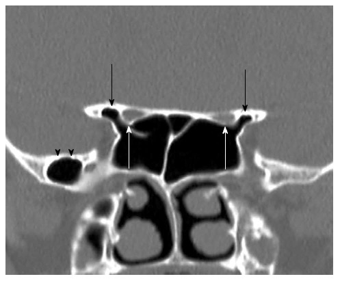
Coronal reformatted computed tomography image shows bilaterally anterior clinoid process pneumatization (black arrows) and optic nerves protrusion (white arrows) into sphenoid sinuses. Additionally shows right greater sphenoid wing pnematization (black arrow heads).