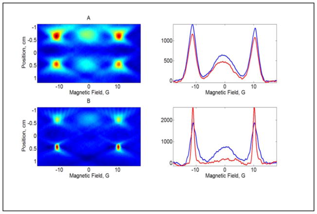
Figure 7.
2D spectral-spatial images of II and IIa in a two-compartment phantom with a 10 mm spacer between compartments. A) Left: both compartments contain 0.5 mM diradical II; right: slices through the upper (blue) and lower (red) compartments of the image. B) Left: the upper compartment contains 0.5 mM II and the lower compartment contains 1 mM IIa, generated by the reaction of 0.5 mM II with 1 mM glutathione; right: slices through the upper (blue) and lower (red) compartments of the image.

