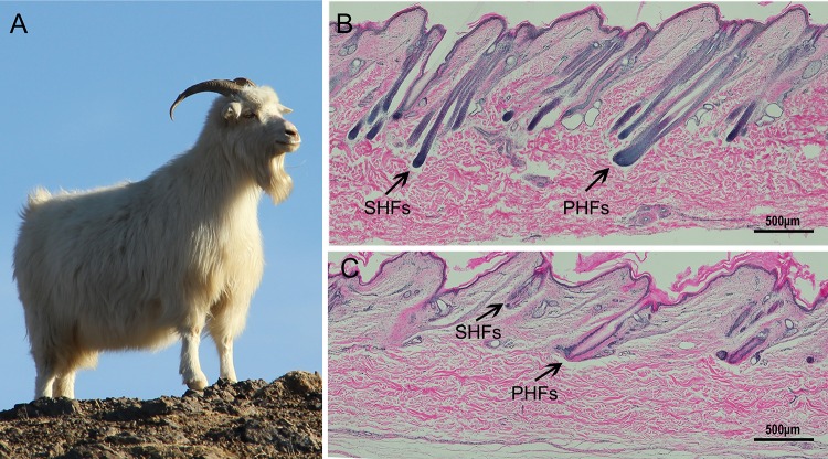
Fig 1. Photograph of white Arbas Cashmere goat and paraffin sections of Cashmere goat skin stained with hematoxylin & eosin.
A. Photograph of a white Arbas Cashmere goat. B. Longitudinal section of goat skin sampled during the short photoperiod (anagen phase). C. Longitudinal section of goat skin sampled during the natural photoperiod (pro-anagen phase). The black arrows indicate the primary hair follicles (PHFs) and secondary hair follicles (SHFs) in the samples. Scale bars: 500 μm.

