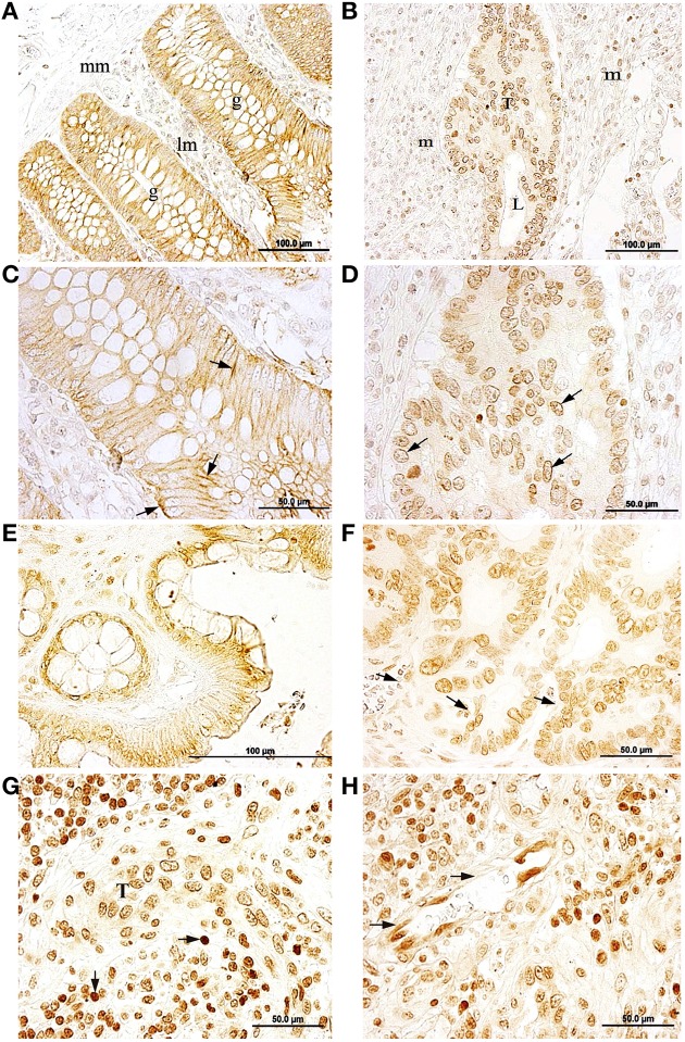
Figure 1.
Immunolocalization of the Na,K-ATPase α1 and α3 subunit isoforms in normal colon and colorectal cancer (CRC) (A,C) α1 immunostaining in baso-lateral side of polarized epithelial cells in healthy colonic mucosae, black arrow. g, colonic gland (Lieberkühn crypts); mm, muscularis mucosae; lm, lamina propia mucosae. (B,D) peri-nuclear labeling for the α1 isoform in tumor cells. T, tumor; L, lumina; m, mesenchymal tissue. (E) α3 immunostaining in plasma membrane of healthy colon epithelial tissue. (F) α3 expressed peri-nuclearly in CRC tumor (arrows). (G) α3 immunostaining in tumor cells (T) surrounded by immune cells (arrows). (H) Endothelial cells expressing the α3 isoform in a peri-nuclear location (arrows).

