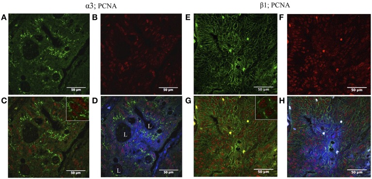
Figure 3.
Double immunofluorescence localization of Na,K-ATPase α3 and β1 isoform and proliferating cell nuclear antigen (PCNA) in CRC. Left panel: (A) α3 isoform is expressed in colon tumor cells (green). (B) High numbers of tumor cells express PCNA (red). (C) The tumor cells express both PCNA and the α3 isoform (blue and green merged). (D) α3 isoform is mainly located internally at the cytoplasm; blue (DAPI), red (PCNA), and green (α3 isoform) merged image. L, lumina. Right panel: (E) β1 isoform is expressed in colonic tumor cells (green). (F) High number of tumor cells expresses PCNA (red). (G) The tumor cells express both PCNA and β1 isoform (red and green merged). (H) Blue (DAPI), red (PCNA), and green (β1 isoform) merged image.

