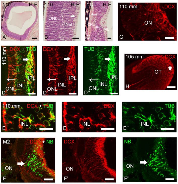
Figure 3.

Photomicrographs of transverse sections of retina and the optic tectum of premetamorphic and metamorphic larval lampreys stained with H-E (A–C) or immunostained for DCX, TUB and/or NB (D–H). (A) Low magnification photomicrograph showing the central and lateral neural retina. Note that the lateral retina extends through most of the retina excepting the central region and a thin irideal retina. Dash lines mark the limit between the central and lateral retinas and between the central and peripheral portions of the lateral retina. (B) Detail of the central portion of the lateral retina showing distinguishable inner (INbL) and outer (ONbL) neuroblastic layers. A thin inner region with differentiated ganglion cells is also observed (thick arrow). (C) Detail of the central retina. The thin arrow points to photoreceptors. The asterisk indicates the dorsal retinal pigment epithelium bearing melanin granules. Dotted lines mark the limits between layers in the lateral retina in (B) and in the central retina in (C). (D–D″) Double immunolabeling for DCX and α-tubulin in the central retina. Note the absence of DCX immunoreactivity in photoreceptors (thin arrow) and its presence in ganglion cells (thick arrow) and in cells of the INL. Note also the DCX/α-tubulin-ir double-labeled radial fibers coursing in the INL and ONL and DCX-ir/α-tubulin-negative end feet in the outer limitans membrane (OLM). (E–E″) Detail of the INL of the central retina of a premetamorphic larva showing the presence of DCX/α-tubulin-ir double-labeled cells. (F–F″) Absence of DCX immunoreactivity in the ganglion cells labeled after application of neurobiotin in the ON of a metamorphic larva (M2). (G) Detail showing DCX-ir fibers in the ON. (H) DCX-ir optic fibers in the optic tectum (asterisk). In all photomicrographs dorsal is up and medial is to the left. (D–F): Overlay; (D′–F′): DCX; (D″,E″): α-tubulin; (F″): NB. Scale bars = 75 μm in (F–F″); 60 μm (A); 50 μm (H); 25 μm (D–E″,G); 10 μm (B,C).
