
Figure 4.
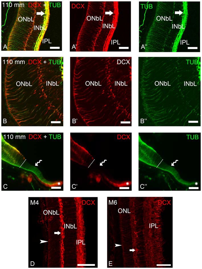
Photomicrographs of transverse sections of the retina of premetamorphic and metamorphic larval sea lampreys showing anti-DCX and anti-α-tubulin immunoreactivities. (A–A″) DCX/α-tubulin-ir double-labeled ganglion cells and fibers in the IPL and cells of the outer layer of the INbL in the lateral retina. Note the presence of double labeled radial fibers coursing throughout the neuroblastic layers and ending in the OLM. (B–B″) Detail of the lateral neural retina of a premetamorphic larva showing the presence of DCX/α-tubulin-ir double-labeled radial fibers in the INbL and ONbL. (C–C″) Photomicrograph of the border of the lateral neural retina of a premetamorphic larva showing the lack of DCX immunoreactivity in the growing irideal retina (curved arrow). Note that the outer epithelium of the irideal marginal region was DCX-ir (asterisks). Dash lines limit the marginal retina from the rest of the lateral retina. (D) In M4 larvae, DCX immunoreactivity was observed in fibers in the IPL, in radial fibers of the INbL and ONbL ending in the OLM (arrowhead) and in cells in the outer INbL (arrow). (E) In M6 larvae, DCX immunoreactivity was similar but retinal layers appear differentiated. (A–C): Overlay; (A′–C′): DCX; (A″–C″): α-tubulin. Scale bars: 50 μm (A–A″,C–C″); 37.5 μm (D,E); 25 μm (B–B″).