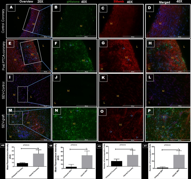
Fig 4. Double immunofluorescence showing expression of pHistone H3 at Ser-10 and SMemb in thin sections of uninjured swine coronaries, post-PTCA coronary arteries, uninjured swine SEV and SEV-graft.
There is less immunopositivity to pHistone (green) and SMemb (red) expression in uninjured coronary artery shown at low (20x) (A) and high (40x) magnification (B, C, D). The immunostaining of pHistone (green) and SMemb (red) is noted in restenotic post PTCA- coronary arteries (arrows) at low (20x) (E) and high (40x) magnification (F, G, H). Similarly, less immunostaining of pHistone (green) and SMemb (red) expression is noted in the uninjured SEV shown at low (20x) (I) and high (40x) magnification (J, K, L) compared to SEV post CABG, where many cells showed immunopositivity to pHistone (green) and SMemb (red) at low (20x) (M) and high (40x) (N,O,P) magnification. This is a representative of tissue histology in 2–5 individual swine in each experimental group. MFI measured in arbitrary units (AU) in post-PTCA coronary arteries (Q) and SEV-grafts (R) compared to their uninjured counterparts. Graphs showing number of pHistone- positive (+) cells in the neointima of the post-PTCA restenotic coronary artery (S) and SEV-grafts (T) compared to their uninjured counterparts. Strong immunopositivity to pHistone H3 was found in the neointima of the post PTCA-coronary arteries and in SEV-graft. Many cells in the neointima of the injured vessels were immunopositive for both, pHistone H3 and SMemb, especially around the pseudo lumen in SEV-graft. Data are shown as mean ± SEM of values of at least three measurements in each group in 2–5 individual swine in each experimental group. *P < 0.05. L: lumen, M: medial layer, NI: neointima.

