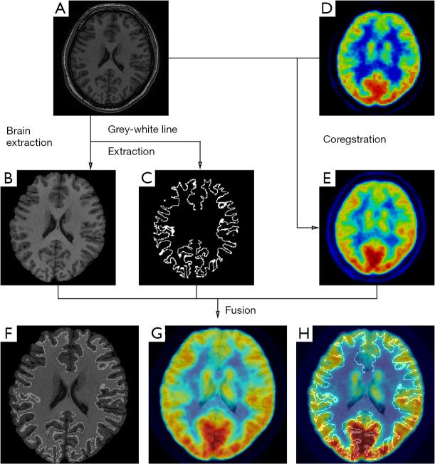
Figure 1.
A 23-year-old female presented with frequency nocturnal hypermotor seizure since she was 2 years old, which was refractory to medical therapy. MRI and FDG-PET image were unremarkable. To detect possible epileptic lesion, MRI-PET coregistration was performed. Based on raw 3D T1 MRI (A), brain (B) was extracted. The junction line of gray and white matter line (C) was also extracted. Meanwhile, raw FDG-PET (E) was coregistered on raw 3DT1 MRI (D). 3D T1 MRI was overlaid with junction line of gray and white matter line (F). On the fused image with MRI and PET (G), slight hypo-metabolism was observed on right anterior cingulate cortex (arrow), but it was likely to be overlooked. However, it revealed mismatch on the same area by overlaid with additional junction line of gray and white matter line, which highlighted the abnormality on the individual gyrus. (Freesurfer and FSL software were used in data processing). Post-operation histological examination showed focal cortical dysplasia IIa. (Unpublished data).

