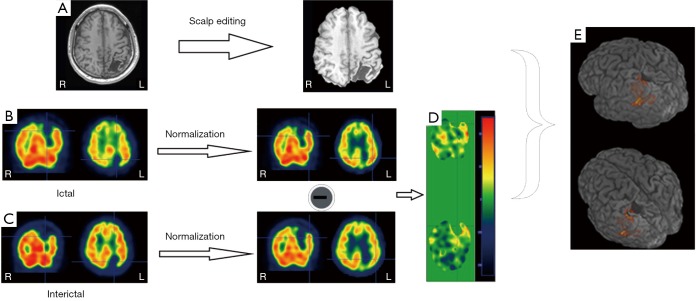
Figure 2.
An 18-year-old male was evaluated epilepsy surgery for refractory seizure that began soon after brain injury at 6 years old. MRI showed large encephalomalacia mainly on left parietal lobe. To delineate the epileptogenic zone, inter-ictal, ictal SPECT and then SISCOM were conducted. Firstly, Raw 3D T1 MRI was processed with skull removal (A). Ictal and inter-ictal 99mTc-SPECT image (B,C) was normalized on normal template followed by the subtracted. Finally, the different image (D) was display on 3D MRI, showing significant hyperfusion on lateral of lesion (3 standard deviations) (E). (SPM software). (Unpublished data).

