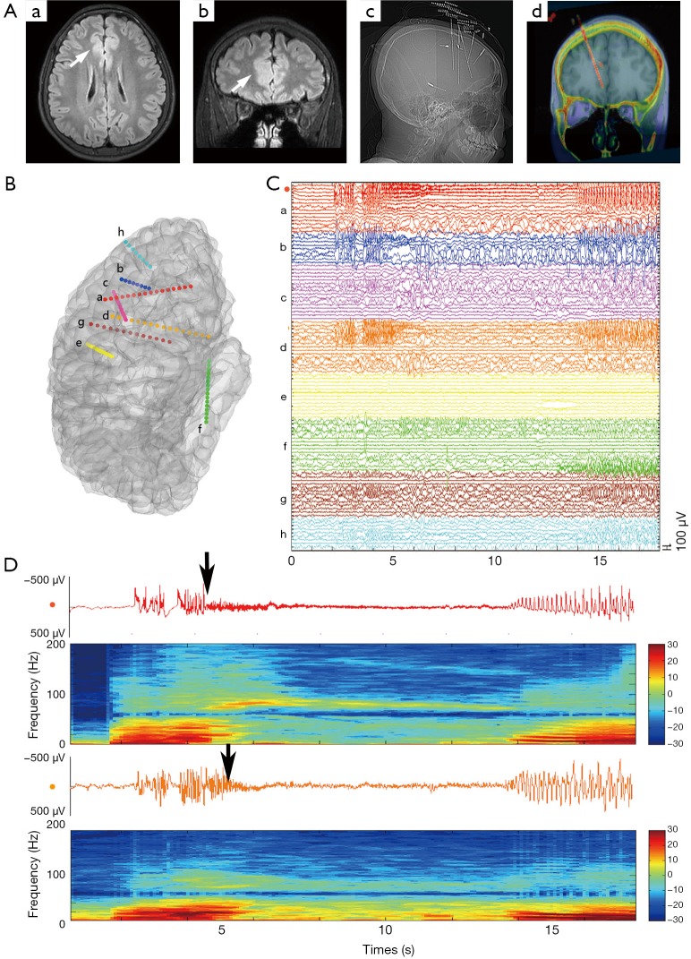
Figure 5.
A 17-year-old male presented with refractory focal seizure since he is 4 years old. Habitual seizure manifested with aura with fear, followed by integrated gestural motor behavior lasting from 10 to 30 s. MRI scan showed subtle signal alteration on right anterior cingulate gyrus (Aa,b). SEEG was implanted based on the assumed seizure onset zone (anterior cingulate gyrus) and possible seizure network involved (Ac). Post-operation CT was coregistered on pre-operation 3D MRI (Ad) to get the spatial location of each electrode precisely. Ictal SEEG epoch and the reconstruction of electrodes and brain were displayed (B). It showed seizure onset from the inner contacts (small numbers) of electrode A rather than outer contacts (large numbers). To highlight the seizure propagation, brain activities on electrode A2 (anterior cingulate cortex, red) and on electrode D2 (obito-frontal lobe, orange) were further demonstrated together with time-frequency analysis. It showed typical seizure onset with fast activity (about 80–100 Hz) on electrode A2 is about 1 s earlier than D2 (arrows). Post-operation histological examination showed focal cortical dysplasia IIa. (Unpublished data).

