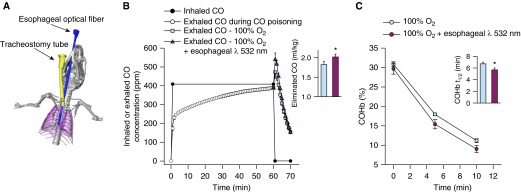
Figure 6.
Esophageal phototherapy increases the rate of carbon monoxide (CO) elimination. (A) Computed tomography scan of a living, anesthetized, and mechanically ventilated 25-g mouse with an optical fiber placed in the esophagus as the three-dimensional reconstruction form. The esophageal optical fiber is depicted in blue, the lungs in purple, and the tracheostomy tube in yellow. (B) Inhaled and exhaled CO concentrations of mice during poisoning with 400 ppm CO (0–60 min) and treatment (60–70 min) by breathing 100% O2 alone or combined with intermittent transesophageal phototherapy at 532 nm (n = 6 per group). (B, inset) The areas under the curves of exhaled CO concentration during the first 5 minutes of treatment were greater in mice treated with 100% O2 and esophageal phototherapy as compared with mice breathing 100% O2 alone (*P < 0.001; Student’s t test). (C) Arterial blood carboxyhemoglobin (COHb) levels decreased more rapidly in the first 10 minutes of treatment with esophageal phototherapy and 100% O2 breathing as compared with breathing 100% O2 alone and (C, inset) COHb half-life (COHb-t1/2) was significantly shorter (*P < 0.001; Student’s t test). All data represent mean ± SD.

