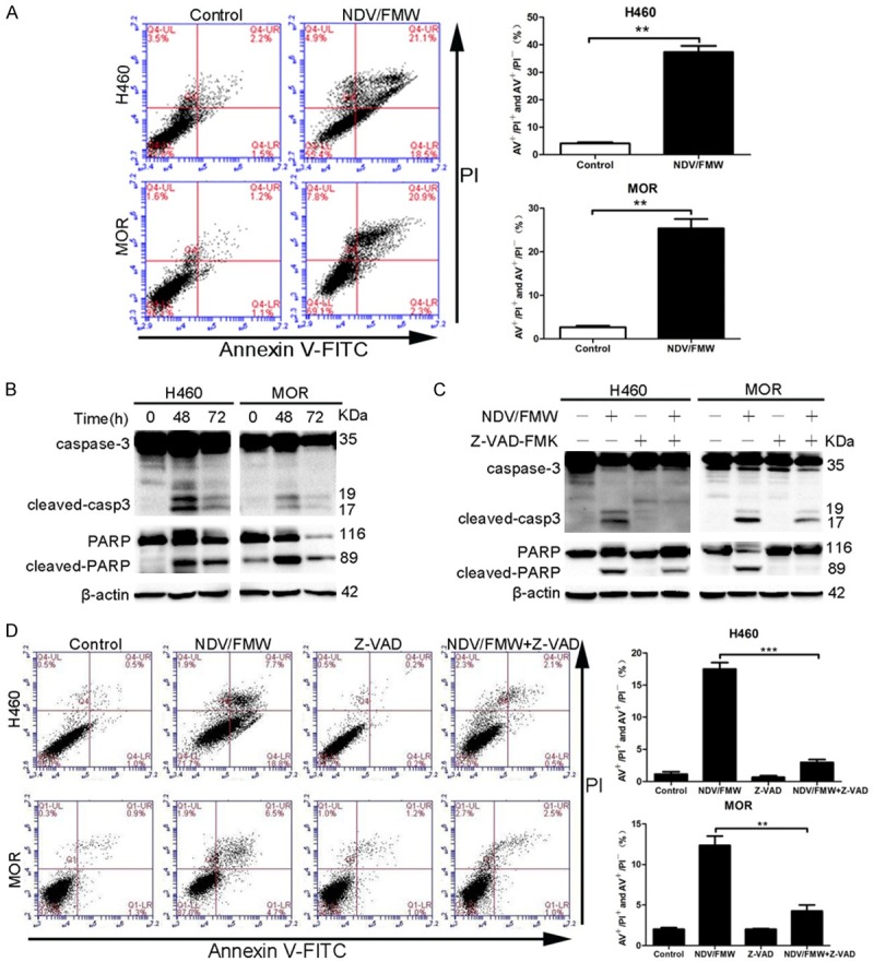
Figure 3.

NDV induces apoptosis of 3D spheroid cultures. A and B. H460 and MOR spheroid cells were mock-treated or infected with NDV/FMW (MOI = 10; 48 h) and double-stained with Annexin V and propidium iodide (PI) and analyzed by flow cytometry. A. The cell population in the right lower quadrant (PI-negative, Annexin V-positive) and the right upper quadrant (Annexin V/PI positive) are represented. Data shown are representative of three independent experiments (**P < 0.01). B. Spheroid cells were mock-infected or infected with NDV/FMW (MOI = 10; 48 and 72 h). Activation of caspase-3 and cleaved poly (ADP-ribose) polymerase (PARP) was examined by immunoblot analysis (n = 2). To control for loading, β-actin was used. C. Spheroid cells were mock-infected or infected with NDV/FMW (MOI = 1; 72 h) in the absence or presence of the pan-caspase inhibitor Z-VAD-FMK (100 μm). Total protein lysates were separated by SDS-PAGE and probed with antibodies against cleaved and total caspase-3 and PARP. All IB experiments were performed twice. D. Apoptosis was also assessed by flow cytometry. The percentage of apoptotic cells are represented as mean ± SD from three independent experiments (**P < 0.01, ***P < 0.001).
