Figure 4.

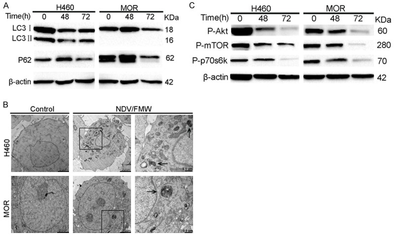
NDV/FMW promotes autophagy flux in 3D spheroid cultures via inhibition of the AKT/mTOR pathway. Spheroid cells were mock-infected or infected with NDV/FMW at an MOI of 10 at the indicated time points. Activation of P62, LC3I to LC3II conversion. (A)And phosphorylated Akt, mTOR and p70S6K were analyzed by immunoblot analysis using β-actin as a loading control. All IB experiments were performed twice (C). Transmission electron microscopy analysis of cells infected with NDV/FMW for 72 h (B).
