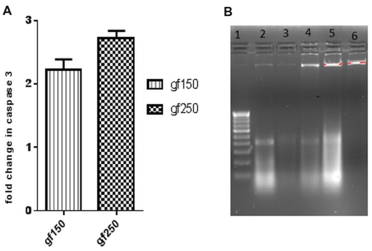
FIGURE 3.
In vitro apoptotic effect of GF on DLA cells was determined by (A) fold change in level of caspase3 of DLA cells on treatment with GF (150 and 250 μg/ml) for 3 h. Fold change is calculated by comparing with untreated DLA cells. All the results are expressed in mean ± SD (n = 3). (B) DNA fragmentation in different doses of GF treated DLA cells. 10 μg DNA from each treatment groups and untreated group was loaded in each lane and subjected to1.8% agarose gel electrophoresis followed by detection of EtBr stained DNA bands in UV transilluminator. The photograph is a representative of three repeats. Here lane1:200 bp DNA ladder, lane2, 3, 4, and 5 represents DNA of DLA cells treated with 50, 100, 150, and 200 μg/ml GF for 3 h. lane6: DNA of untreated DLA cells.

