
Figure 1.
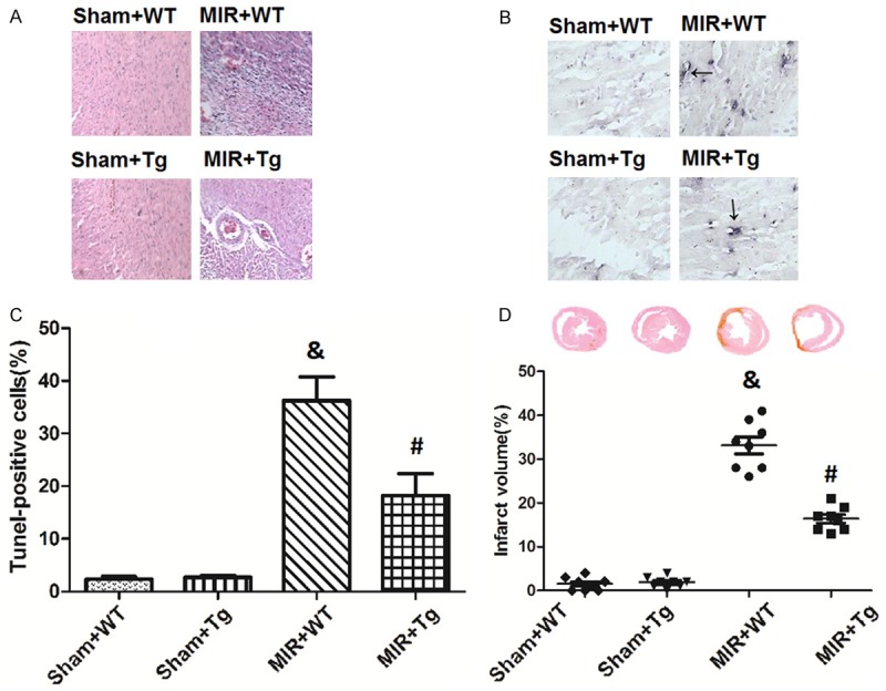
Histopathological, apoptotic and infarct size changes by HSPA12B. Microscopic examinations of the myocardium tissues were stained with hematoxylin and eosin (A). Cellular apoptosis were evaluated via TUNEL staining (B and C). Myocardium infarction was stained with Sirius Red (D). Values are expressed as the Mean ± SD. &p<0.05 compared to the Sham+WT; #p<0.05 compared to the MIR+WT. N = 8 per group.