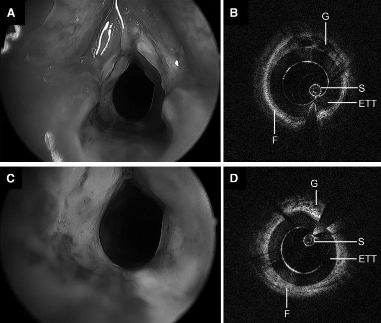
Figure 9.
Operative bronchoscopy and long-range optical coherence tomography images of a neonatal airway following 42 days of intubation, including two failed extubation attempts. Significant edema and granulation, circumferential laryngeal stenosis (A), and grade 2 subglottic stenosis (C) are noted on endoscopy. Long-range optical coherence tomography of the larynx (B) and subglottis (D) demonstrates a thick, hyperintense submucosa with focal patches of hypointensity. ETT = endotracheal tube; F = fibrosis; G = glandular structures; S = sheath.

