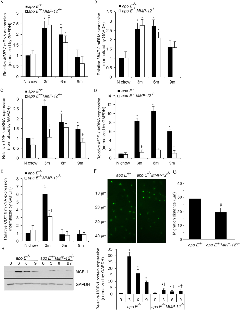
Figure 2. MMP-12 deletion suppresses HFD-induced profibrotic and proinflammatory gene or protein expression in isolated renal glomeruli correlation with macrophage invasion.
(A–E) The mRNA expression levels of MMP-2 and -9, TGF-β, MCP-1, and CD11b were evaluated by qRT-PCR. (F–G) Macrophage cells were seeded on the top of collgen IV gel and after 48 hours were fixed and stained by CD68 (green). The images represent vertical position of optical sections captured at 10 μm intervals from top of matrix (0 μm) to 40 μm of invasion depth. (H,I) Representative western blot (H) and quantification (I) of relative MCP-1 protein levels in apo E−/− and apoE−/−MMP-12−/− mice fed normal chow or HFD for 3, 6, and 9 months. Values represent means ± SEM (n = 6 per group). *P < 0.05 vs. mice on normal chow; †P <0.05 vs. mice on HFD at the same time point; #P < 0.05 vs. apo E−/− mice.

