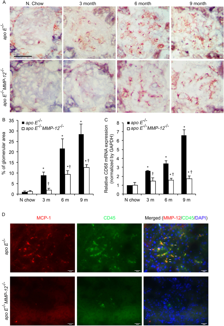
Figure 4. MMP-12 deletion suppresses HFD-induced macrophage infiltration into renal glomeruli.
(A) Expression of the macrophage marker CD68 in apo E−/− and apo E−/−MMP-12−/− mice fed normal chow or HFD for 3, 6, and 9 months. Scale bar = 20 μm. (B) Quantitative analysis of glomerular CD68 expression calculated as a percentage of positive staining within the glomerular area (10 glomeruli per kidney per animal, n = 12 per group). (C) HFD induced CD68 mRNA expression in isolated renal glomeruli evaluated by qRT-PCR. Values represent means ± SEM (n = 6 in each group). *P < 0.05 vs. mice on normal chow; †P < 0.05 vs. mice on HFD at the same time point. (D) Co-localization of MCP-1 and CD45 in renal glomeruli. Immunofluorescent staining show MCP-1 (red) and CD45 (green) in tissue sections from in apo E−/− and apo E−/−MMP-12−/− mice fed HFD for 3 months. The co-expression of MCP-1 in CD45+ cells was observed in apo E−/− mice glomeruli (arrow). Nuclei are shown counterstained with DAPI (blue). Scale bar = 20 μm.

