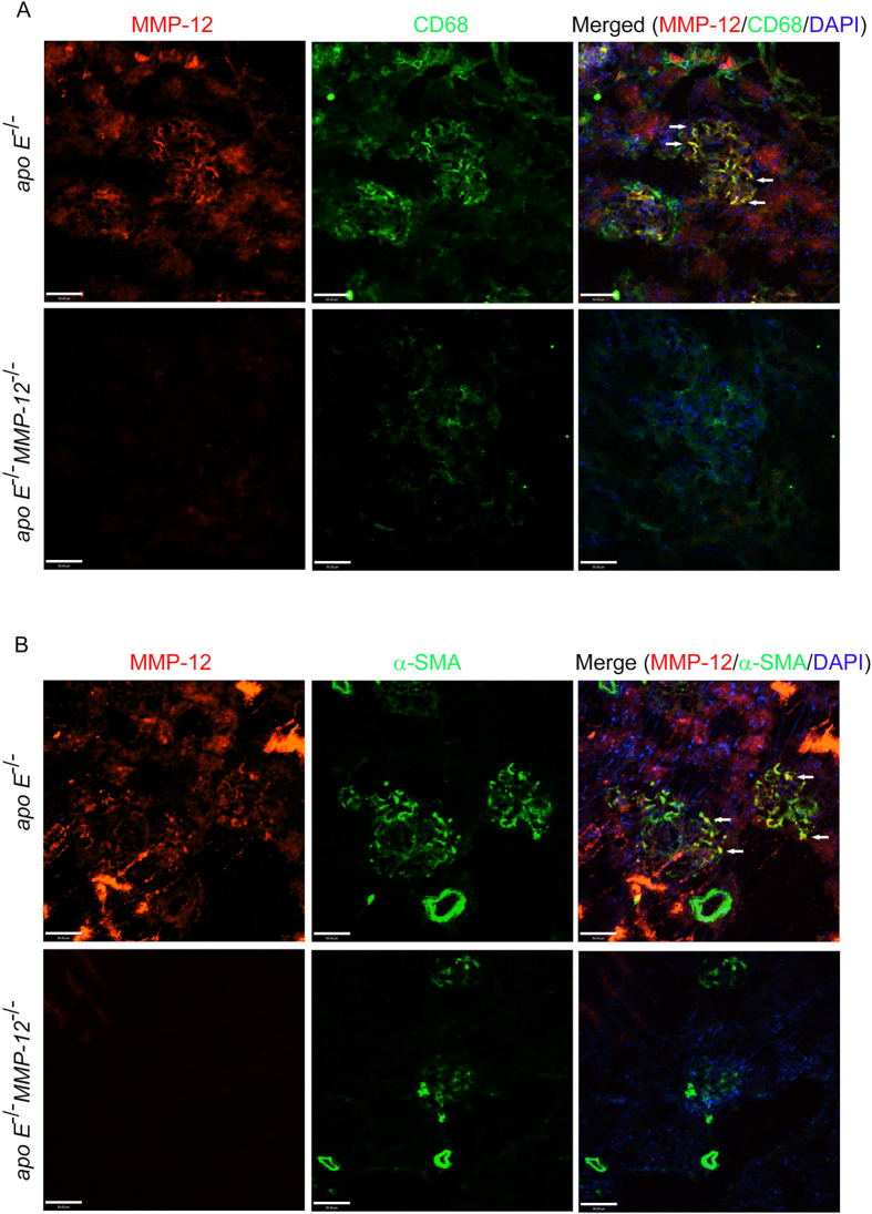
Figure 5. Co-localization of MMP-12 and CD68 or α-SMA in renal glomeruli.
(A) Confocal images of MMP-12 (red) and CD68 (green) immunoreactivity in tissue sections from apo E−/− and apo E−/−MMP-12−/− mice maintained on HFD for 6 months. Most CD68+ infiltrated macrophages coexpressed MMP-12 in apo E−/− but not apo E−/−MMP-12−/− mice (arrow). Nuclei were counterstained with DAPI. Scale bar = 50 μm. (B) Co-localization of MMP-12 and α-SMA expression in renal glomeruli. Confocal images show MMP-12 (red) and α-SMA (green) immunoreactivity in tissue sections from apo E−/− and apo E−/−MMP-12−/− mice fed HFD for 6 months. The co-expression of MMP-12 in α-SMA+ mesangial cells was observed in apo E−/− glomeruli (arrow). Nuclei are shown counterstained with DAPI (blue). Scale bar = 50 μm.

