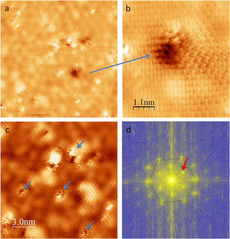
Figure 2. STM images of fluorinated graphene (FG).
(a) 20 × 20 nm2 area, (b) Zoom in image of a hole defect showing standing waves pattern, (c) Other area 15 × 15 nm2 showing bright feature decorating holes (blue arrows) attributed to fluorine atoms. (d) shows the FFT (Fast Fourier Transform) of (a). This reveals the first Brillouin zone with the hexagonal lattice and K points (red arrows) associated to the standing waves pattern due to intervalley scattering.

