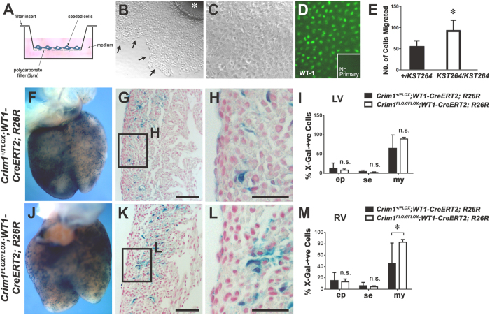
Figure 4. A cell-autonomous requirement for Crim1 to control epicardial migration and myocardial invasion in vitro and in vivo.
(A) Schematic of the transwell assay used for the in vitro migration assay. (B) Representative micrograph of a primary 11.5 dpc ventricular explant after attachment and migration of epicardial cells. The front of the epicardial monolayer is indicated (arrows). The primary explant is denoted by an asterisk. (C) Primary epicardial cells after removal of the ventricular explant, passage and subsequent culture. Note maintenance of a “cobblestone” appearance of the cellular monolayer. (D) Epicardial identity after enrichment was confirmed by immunostaining for WT1 (green nuclear signal). The no-primary antibody control showed no signal (inset). (E) Crim1KST264/KST264 primary epicardial cells showed increased migration in vitro relative to controls (n = 3–5 and representative of three independent experiments; *P < 0.05). (F,J) Whole-mount dorsal views of 15.5 dpc embryonic hearts after X-Gal staining (blue) of Crim1+/FLOX; WT1-CreERT2; R26R and Crim1FLOX/FLOX; WT1-CreERT2; R26R genotype, respectively. Two doses of Tamoxifen were administered at 9.5 dpc and 10.5 dpc prior to harvesting the embryos. (G,K) Histological sections of hearts from Crim1+/FLOX; WT1-CreERT2; R26R and Crim1FLOX/FLOX; WT1-CreERT2; R26R 15.5 dpc embryos, respectively, and (H,L) magnified views of boxed regions showing X-Gal-positive epicardial, sub-epicardial and intramural cells. (I,M) Quantification of X-Gal-positive cells showing increased myocardial EPDCs in the right ventricles of mutant hearts (n = 4–6; *P < 0.05). ep, epicardial; se, sub-epicardial; my, intramyocardial; LV, left ventricle; RV, right ventricle. Scale bars, (G,K) 20 μm (H,L) 10 μm.

