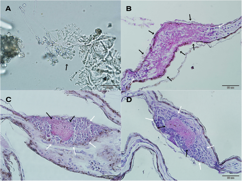
Figure 2. White-nose syndrome on a pond bat Myotis dasycneme near Yekaterinburg, Russia, in May 2014.
(A) Microscopic identification of the characteristic curved conidia of Pseudogymnoascus destructans (black arrow). (B) Invasive fungal growth penetrating the full-thickness of the wing membrane (black arrows), with several inflammatory cells (neutrophils) situated at both margins of the lesion (white arrows). (C) Packed fungal hyphae of cupping erosions (black arrows) sequestered by neutrophils (white arrows). (D) Histopathological finding from a WNS-positive Nearctic Myotis lucifugus, identical to that found in Palearctic Asia. Skin sections stained with periodic acid-Schiff stain.

