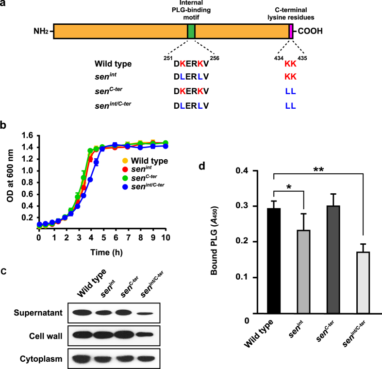
Figure 4. Introduction of sorting signal into SEN affects subcellular localization and bacterilal PLG binding.
(a) Schematic diagram of SEN protein. Internal and C-terminus PLG-binding motifs are shown as green and pink images, respectively. The amino acid sequences of the PLG-binding motif in each strain are described below. (b) NIH35 and isogenic sen mutants were grown in THY broth. The culture densities were measured at 37 °C. (c) The strains were grown to an OD600 of 0.8 in THY broth, then each fraction was prepared, as described in Methods. (d) NIH35 and the isogenic sen mutants (OD600 = 0.6) were bound to microtiter plates, and bound cells were incubated with 1 μM human PLG. Cell-bound PLG was detected by ELISA using an anti-PLG antibody. Data are shown as the mean ± S.D. of six samples from a representative experiment. *P < 0.05; **P < 0.01.

