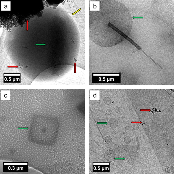
Figure 2.

Bright field TEM images of SSA prepared by cryo-TEM showing (a) a whole bacterium inside a wet SSA droplet (note: the image contrast and brightness was adjusted to aid the observation of the cell, the original image is shown in Figure S11), (b) an intact diatom, (c) a virus particle, and (d) marine membrane vesicles. The biological structures were identified according to their size, shape, and morphology (see Supporting Information for more details). Yellow arrows indicate the edge of the SSA, the red arrows indicate contamination from the cryo-TEM preparation, and the green arrows indicate the biological particles.
