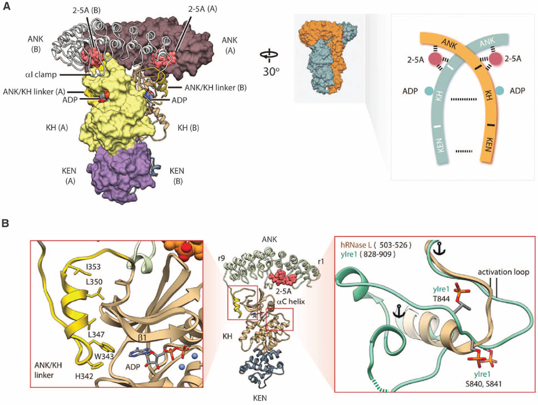
Fig. 1. Structure of human RNase L•2–5pA3•ADP•RNA18 quaternary complex.
(A) Crossed homodimer of RNase L. Different protomers are shown as molecular surface and ribbon, respectively. The structured linker connecting the ANK and the KH domains is colored gold. Schematic topology of the RNase L homodimer is shown on the right. (B) Structure of the KH domain. The ATP pocket is flanked by the structured linker (left). The activation loop is short and lacks phosphorylation sites (right). Single-letter abbreviations for the amino acid residues are as follows: A, Ala; C, Cys;D, Asp; E, Glu; F, Phe; G, Gly; H, His; I, Ile; K, Lys; L, Leu; M, Met; N, Asn; P, Pro; Q, Gln; R, Arg; S, Ser; T, Thr; V, Val; W, Trp; and Y, Tyr.

