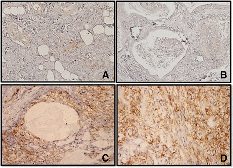
Fig. 1.

Immunohistochemistry for CD44 in Mucoepidermoid carcinomas. a Normal salivary glands showed weak cytoplasmic stain in ductal epithelium and no immunoreactivity is seen in acinic cells. b No or mild CD44 expression was observed in the membrane of mucoepidermoid carcinoma cells (low grade type). c, d CD44 expression was strongly expressed in the membrane of the mucoepidermoid carcinoma cells (intermediate and high grade type)
